The long reach of the Hox code
LMU researchers have shown that patterns of activity of certain genes in vertebrate embryos correlate with differences in the length of the neck region. The findings also provide new insight into the evolution of long-necked dinosaurs.
17.06.2015
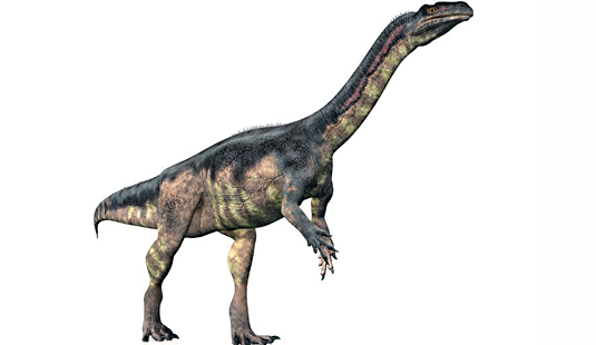
Plateosaurus (Source: Michael Rosskothen / Fotolia.com)
Crocodiles, birds – and dinosaurs – are closely related to each other, and all are included within a larger evolutionary lineage called archosaurs. In some archosaurs, such as long-necked dinosaurs and modern birds, the number and disposition of the vertebrae in the vertebral column is extremely variable. One of the most striking features of avian evolution – exemplified by the ostrich but also found in the long-necked dinosaurs – is an increase in the number of cervical vertebrae. How this variation in the structure of the anterior vertebral column arose is not at all clear. “Particularly in the context of dinosaur evolution, there has been a great deal of discussion as to whether these changes result from displacements of the different regions of the axial skeleton or are due to the insertion of novel elements,” says Christine Böhmer of LMU’s Department of Earth and Environmental Sciences and the Bavarian State Collection for Palaeontology and Geology. In collaboration with her Munich colleagues Oliver Rauhut and Gert Wörheide, Böhmer has asked whether changes in the structure of the cervical vertebrae in present-day archosaurs are reflected in the activity of a crucial set of developmentally important genes called Hox genes.
Hox genes define morphological borders
The major function of the Hox genes in vertebrates is to choreograph axial patterning during embryonic development. They play a central role in the spatial and temporal regulation of genes required for the formation of the different types of vertebrae found in different regions of the axial skeleton. Specific Hox genes are activated only at certain times and in specific segments of the embryo during early development, and their expression patterns can be visualized using molecular biological methods. These experiments reveal characteristic expression patterns for each Hox gene, which show up as a defined sequence of stripes along the anterior-posterior axis of the growing embryo. “It was already known that borders of some of these ‘expression domains’ correspond to morphological boundaries in the vertebral column – for example, that between cervical and dorsal vertebrae: Formation of the two groups of vertebrae requires the activation of different subsets of Hox genes,” Böhmer explains.
Böhmer and her colleagues studied the expression patterns of various Hox genes during early development in crocodiles and birds. The results showed that limits of Hox expression domains not only demarcate clearly defined regions of the vertebral column – cervical and dorsal, for instance – but that the margins of subdomains reflected morphological differences between vertebrae within these regions. “The crocodile has four units of cervical vertebrae, while the neck of the chicken contains five. Accordingly, four and five Hox expression domains, respectively, are found in developing crocodile and chick embryos, and their borders coincide with those between the different vertebrae. The pattern in the crocodile mirrors the ancestral configuration. During the course of evolution, this pattern was altered in such a way that the neck region got longer,” Böhmer adds.
Crocodile – dinosaur – chick
The Munich researchers then took a closer look at the morphology of the cervical vertebrae in the long-necked dinosaur Plateosaurus, of which several well-preserved fossil specimens are available. “We found four morphologically distinct subdomains within the cervical vertebral column, as in the crocodile. Strikingly, however, there are two vertebrae in the second subdomain, as in the chick,” Böhmer explains. This strongly suggests that an increase in the number of vertebrae in the second sub-region of the ancestral cervical column initiated the process that led to the extended neck region seen in modern-day birds. “And that most probably resulted from a change in the pattern of Hox gene expression,” says Böhmer. “Our findings argue that the second step in the elongation of the neck involved the emergence of a new sub-region boundary and a corresponding change in genetic expression patterns, like the one we see in the chick.”
The team now plans to investigate other fossils with a view to reconstructing Hox expression patterns along the anterior-posterior axis on the basis of regional differences in the morphology of their vertebrae. “That would allow us to infer the nature of the mechanisms that led to changes in vertebral structures, and would yield new insights into the genetic basis of evolutionary change – even in extinct groups like the dinosaurs, for which direct genetic evidence is inaccessible,” Böhmer concludes.