The secrets of the brachiopod shell
LMU researchers have carried out the first detailed study of the molecular mechanisms responsible for formation of the brachiopod shell. Comparison with shell synthesis in other groups reveals the deep evolutionary roots of the process.
04.05.2015
 Brachiopods (lamp shells) are marine invertebrates, which were a highly successful and widespread group in the Palaeozoic era. Indeed, the group is best known for its rich fossil record. Some 30,000 fossil species have been described so far, and the oldest specimens date from Cambrian times, and are thus around 500 million years old. Brachiopods are now represented by comparatively few species, which are found in various regions of the world’s oceans. One of their most characteristic features is their bipartite shell. “The molecular basis of how this shell is actually built has been virtually unknown up to now,” says Professor Gert Wörheide of the Department of Earth and Environmental Sciences and the Geobio-Center at LMU. “To find out more, we have carried out the first comprehensive survey of the genes and proteins involved in shell formation in brachiopods”. The results of the study appear in the journal “Genome Biology and Evolution”.
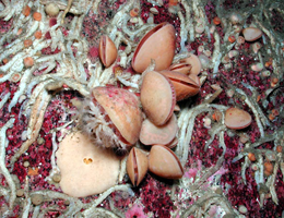
Although brachiopods look very much like mollusks at first sight, they are not related to the latter. In contrast to the typical mollusk shell, their bipartite shell is not left/right symmetrical. Instead it comprises an upper (dorsal) part and a lower (ventral) ‘valve’, with the ventral valve usually being the larger. The valves are made of either calcium carbonate (calcite) or calcium phosphate in association with a diverse array of proteins and polysaccharides, which are secreted by the cells of the underlying mantle during shell formation and are incorporated into the biomineral formed. “This is why the proteins occluded within the shell can provide insights into its mode of formation,” Wörheide explains.
To gain such insights, the researchers identified the complete set of proteins (the proteome) found in the shell of the South American brachiopod Magellania venosa. This enabled them to then characterize the genes that encode the blueprints for synthesis of the proteins from the mantle tissue secreting the shell (the mantle transcriptome). “This is the first time that such a screening approach has been applied to any species of brachiopod, and it was the combination of the two methods that allowed us to identify the molecular components involved in formation of the shell,” Wörheide points out.
The results of these two types of molecular screen are of particular interest when compared with data from other groups of shell-forming organisms, such as corals, sea urchins and mollusks. The seven most abundant proteins found in the Magellania shell turn out to be unique to brachiopods, but they exhibit biochemical features similar to those of proteins that are known to serve similar functions in other animal phyla. Other proteins show significant structural resemblances to shell proteins that are known from other animals. Based on these analyses, the researchers conclude that the genetic program and the molecular mechanisms utilized in biomineralization – the biochemical processes by which living organisms synthesize mineral-based structures in a controlled manner – have, in part, been evolutionarily conserved among invertebrates. “Our results provide entirely new insights into the evolution of shell formation in the brachiopods,” Wörheide says, “and these data will also be very useful in future studies.”
Genome Biology and Evolution 2015